Vous êtes ici : UFR Simone Veil - santéFRFormation continueSeptic shock02. Look for severe LV systolic dysfunction
- Partager cette page :
- Version PDF
02. Look for severe LV systolic dysfunction
Left ventricular systolic dysfunction is classically reported in septic shock. However, in contrast to the opinion of most intensivists, it is not rare. Even if the so-called hyperkinetic profile is predominant.
TEE in a female patient on assisted ventilation because of septic shock due to pneumonia. After filling, the long-axis and short-axis views of the left ventricle indicated marked ventricular hyperkinesis. This often necessitates infusion of high doses of noradrenaline. The prognosis is often gloomy.
We have found a hypokinetic state in 35% of almost 200 patients with septic shock. We define a hypokinetic profile as a combination of a cardiac index below 3 l/min/m2 and of an LV ejection fraction (LVEF) below 40%.
TEE in a patient ventilated for septic shock with severe systolic dysfunction of the left ventricle. Note that the LV did not seem dilated and that its hypokinesis was global. In this situation, it was essential to infuse an inotropic agent.
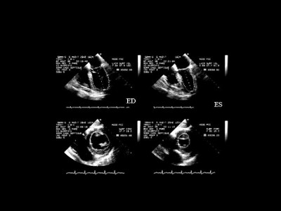
So, LV systolic dysfunction should always be sought. This requires long-axis and short-axis views of the LV, ideally by transesophageal electrocardiography (TEE), but also sometimes by transthoracic electrocardiography (TTE). The long-axis view enables measurement of ventricular volumes and therefore of LVEF (LVEF = end-diastolic volume – end-systolic volume / end-diastolic volume).
The short-axis view allows measurement of ventricular areas and therefore of the LV fractional area contraction, close to its ejection fraction (FAC = end-diastolic area - end-systolic area / end-diastolic area).
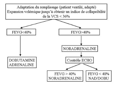
Our therapeutic approach is systematized from echocardiography: in the presence of circulatory insufficiency, or of metabolic acidosis reflecting tissue hypoxia, an LVEF or an LVFAC below 40% systematically requires infusion of an inotropic agent (dobutamine usually, sometimes epinephrine).
Echocardiography must always be repeated after initiation of infusion, to check treatment efficacy.
Septic shock of urinary origin in a female patient on assisted ventilation. TEE revealed global hypokinesis of the left ventricle with an ejection fraction below 40%. Circulatory insufficiency in this patient necessitated infusion of an inotropic agent.
In the same female patient as in film 11, infusion of dobutamine restored left ventricular systolic function and systemic blood pressure.
PSystematic and regular echocardiography should be used to monitor left ventricular function on initiation of treatment with a pure vasoconstrictor like norepinephrine. We have already observed that norepinephrine, by increasing afterload on the LV, can unmask LV systolic dysfunction requiring addition of an inotropic agent.
LV: left ventricle; LA: left atrium; RV: right ventricle; RA: right atrium
Female patient on assisted ventilation for septic shock of urinary origin requiring placement of a urethral probe. In the immediate postoperative period, TEE detected hyperkinesis of the left ventricle. Infusion of noradrenaline was started because of circulatory insufficiency.
LV: left ventricle; LA: left atrium; RV: right ventricle; RA: right atrium
In the same female patient as in Film 13, infusion of noradrenaline revealed, a few hours later, myocardial incompetence of the left ventricle associated with persistence of metabolic acidosis requiring infusion of dobutamine.
LV: left ventricle; LA: left atrium; RV: right ventricle; RA: right atrium
In the same female patient as in Films 13 and 14, the infusion of dobutamine restored left ventricular systolic function and corrected metabolic acidosis.
Media
Figure 3 : Measurement of volumes and left ventricular area by TEE.
ED: end-diastolic, ES: end-systolic
Figure 4 : Protocol for hemodynamic management of septic shock using TEE.
LVEF : left ventricular ejection fraction. NEP : norepinephrine. Dobu : dobutamine.


