Aller au contenu

| | |

Vous êtes ici : UFR Simone Veil - santéFRFormation continueCardiac tamponade01. Pathophysiological aspects01.1 Atrioventricular competition

01.1 Atrioventricular competition

Provided they remain within their physiological limits, distension of the atria and ventricles is not limited by a healthy pericardial space. The sum of the chamber volumes is greater in diastole than in systole. When a pathological effusion occurs, atrioventricular competition is such that the sum of the chamber volumes is constant throughout the cardiac cycle. When the compression is greater, this constant is smaller.

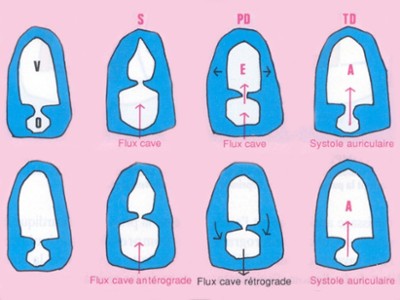
In a physiological setting, sudden reduction in ventricular dimensions at the start of systole creates pericardial depression which drives venous return, and the atria fill by anterograde systolic acceleration of the vena caval flow. The ventricle dilates on diastole and the ventricular depression thus created aspirates the blood of the atrium towards the ventricle (E-wave of rapid filling), but also from the vena cavae towards the atrium, inducing anterograde diastolic acceleration of caval flow. At end-diastole, atrial systole completes ventricular filling (A-wave).


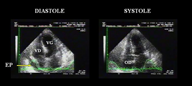
In the pathological setting of CT (Figure 2, Film 1), the atrium can only be filled if the ventricle empties, and vice versa. On systole, the pericardial depression created by the reduction in ventricular dimensions enables atrial filling, and the anterograde systolic acceleration of caval flow is preserved. From the start of diastole, the increase in ventricular volume raises pericardial pressure and atrial compression. The atrium empties towards the zone of least resistance, extracardiac and retrograde: the E-wave of rapid filling is greatly reduced and there is a retrograde diastolic acceleration of caval flow and diastolic atrial collapse (Film 2). 
 

Film 1: Atrioventricular competition in a patient presenting cardiac tamponade. Note on this apical four-chamber view that the right atrium can only be filled when the right ventricle empties.


Film 2: Right atrial collapse in diastole during cardiac tamponade, observed by the subcostal route EP: pericardial effusion; OD: right atrium

At end-diastole, active atrial contraction supplies ventricular filling: the A-wave becomes predominant.

Media

Aucune page à faire défiler
Page suivante
Page précédente
Aucune page à faire défiler
Figure 1 : In the normal physiological situation (upper part of the figure, V: ventricle, A: atrium), the reduction of ventricular volume on systole (S) lowers the pericardial pressure and facilitates atrial filling by anterograde accentuation of caval flow (systolic filling wave, see Fig. 18). In protodiastole (PD), ventricular dilatation raises the pericardial pressure, but the aspiration effect created by this dilatation is greater and favors rapid filling (E) (On the recording of the atrial pressure curve, this aspiration effect is responsible for the hollow y.) At the same time, the atrium is filled by new anterograde acceleration of caval flow (diastolic filling wave, see Fig. 18). In end-diastole (TD), ventricular filling is completed by atrial systole (SA).

During cardiac tamponade (lower part of the figure), the sudden rise in pericardial pressure in protodiastole compresses the atrium and reverses the caval flow (note that the hollow y is missing on a recording of atrial pressure, and that on a recording of caval flow there is no diastolic wave, see Fig. 18). In end-diastole, only atrial systole participates in ventricular filling. The catastrophic consequences of atrial fibrillation in this situation are easy to imagine.

Figure 2 : An apical four-chamber view can be used to observe the pushing in of the atrial wall by the pericardial effusion (EP) in diastole, and the re-expansion of the right atrium (OD) in systole. VD: right ventricle, VG: left ventricle.