Aller au contenu

| | |

Vous êtes ici : UFR Simone Veil - santéFRFormation continueCardiac tamponade01. Pathophysiological aspects01.4 Ventricular interaction

01.4 Ventricular interaction

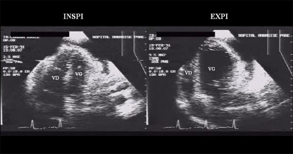
This is another aspect of the ventricular-ventricular competition caused by the restriction of the pericardial space. Inspiration, which steepens the gradient favorable to venous return fed by extrathoracic blood vessels, encourages expansion of the right ventricle. As this expansion cannot occur through encroachment on the pericardial space, it happens through septal shift towards the left ventricular chamber (Figure 7).


Film 4: Apical four-chamber view showing the right (RV) and left (LV) ventricles.
In this patient with cardiac tamponade, inspiration is responsible for improved filling of the right ventricle and restriction of the left ventricle, visualized by movement of the interventricular septum towards the left ventricle. The opposite phenomenon is seen on expiration.

Film 5 : In the same patient as in film 4, the small-axis view of the left ventricle shows the same phenomenon. RV: right ventricle, LV: left ventricle.

The dimensions and distensibility of the left ventricular chamber are thus reduced, which blocks pulmonary venous return. Expiration on the other hand cancels the gradient favorable to venous return, which is therefore interrupted. The resulting reduction in size of the right ventricle allows the septum to return to its place, and the pulmonary venous return is unblocked, permitting filling of the left ventricle.

Media

Figure 6 : On inspiration, pleural depression favors systemic venous return (RVS) and right ventricular filling. Because of a lack of room in the pericardial space, increase in right ventricular diastolic volume shifts the septum leftwards and reduces the diastolic dimensions of the left ventricle. The venous pulmonary pressure increases, and the pulmonary venous return (RVP) is blocked, which leads to blood storage in the pulmonary circulation. On expiration, the systemic venous return is stopped, the septum returns to its place, the pulmonary venous return is restored, and the pulmonary venous circulation empties into the left ventricle. 

Figure 7 : On this apical four-chamber view, note the variations in size of the right (VD) and left (VG) ventricles on inspiration and expiration.