Aller au contenu

| | |

Vous êtes ici : UFR Simone Veil - santéFRFormation continueAcute Cor Pulmonale02. The main echocardiographic findings useful for detecting APC

02. Principal echocardiographic views used to study detect ACP

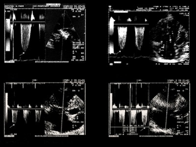
Echocardiographic examination of the RV requires a long-axis view to measure chamber size, and a small-axis view to evaluate the shape and movements of the interventricular septum. To these should be added a view of the right ventricular outflow tract used to assess the Doppler velocity and morphology of pulmonary artery flow, and a Doppler recording of tricuspid flow to detect tricuspid regurgitation and so measure the transtricuspid gradient and deduce from it the pulmonary artery systolicpressure.

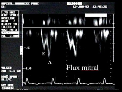
Lastly, it is necessary to examine mitral flow velocity to identify any abnormal left ventricular relaxation.

All these effects and the respective views used to examine them are detailed in the section on the principal echocardiographic views.

When a patient is breathing spontaneously, as is usually the case in pulmonary embolism, the echocardiographic examination is done by the transthoracic route (transthoracic echocardiography, or TTE). In a patient on assisted ventilation, transesophageal echocardiography (TEE) is easy and should therefore be used as it gives better images. This is how ARDS patients are examined.

Echocardiography is above all a qualitative procedure. However, quantitative measurements can also be made during or after acquisition of sequences. Table 1 gives the principal normal values of our laboratory.

Media

 
Aucune page à faire défiler
Page suivante
Page précédente
Aucune page à faire défiler

Figure 3 : Four recordings of tricuspid regurgitant flow. Doppler measurement of the peak velocity (V, arrow) is used to calculate the transtricuspid pressure gradient, which is equal to 4V 2 . If we know the right atrial pressure, or the central venous pressure (CVP), it can be added to this gradient to give the right ventricular systolic pressure. If the exact CVP value is not known, it can be estimated from the measurement of the diameter of the inferior vena cava (diam) at the end of expiration ( CVP= 0.64diam + 0.77). 

Figure 4 :  Abnormal left ventricular relaxation. Abnormal septal motion and compression of the left ventricular chamber caused by right ventricular dilatation are seen in impaired left ventricular filling, which is characteristic of abnormal relaxation: the E-wave, of rapid filling, is reduced, whereas the A-wave linked to atrial systole, is preeminent.